Ultrasound Services
Ultrasounds are one of our common diagnostic procedures that take a greater look at the soft tissues in the body, such as the muscles, organs and blood vessels. Ultrasounds are also non-invasive, meaning the skin is not pierced in order to perform the procedure.
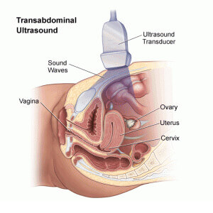
During an ultrasound, a transducer is used to send out ultrasonic sound waves. These sound waves are so high in frequency that they can’t even be heard by the human ear. Once the transducer is placed at a certain angle or location, the sound waves will bounce off the internal organs and echo back to the transducer. The transducer then takes note of the echoed sound waves and coverts them through the help of a computer. This computer will reflect on the waves and create an electronic picture of the organs and tissues.
 Since different body tissues affect the way that sound waves travel in terms of speed, it is easy for the transducer to distinguish between bone tissue and air in the body. Sound waves travel the quickest through bone tissue, while the sound waves travel the slowest through air.
In order for the transducer to move smoothly across the skin, a clear conducting gel must be applied to the skin’s surface. This gel also decreases the chances of air getting between the skin and transducer, which could affect the sound conduction.
In order for the blood flow of a patient to be assessed, a technologist can apply additional ultrasound technology equipment. In order for the ultrasound to assess blood flow, the transducer must contain a Doppler probe. A transducer that contains a Doppler probe will evaluate the direction of the blood flow in the vessels, as well as the velocity by providing audible sound waves. Typically, the louder the sound waves are the more blood flow within the blood vessel. On the other hand, if the sound waves are very faint, that may be a clear indicator that the blood flow is obstructed or blocked.
An ultrasound is one of the best forms of diagnostic care since an ultrasound provides the technologist with the real time functioning of the organs and blood vessels. An ultrasound sound can be used to examine any part of the body. Generally, technologists examine the breasts, abdomen, female pelvis, prostate, scrotum, thyroid and parathyroid glands, as well as the vascular system. With the help of an ultrasound, an obstetrician can evaluate a pregnant mother’s fetus and its development.
Due to advances in technology, ultrasounds can now provide patients and technologists with three-dimensional views, as well as four-dimensional views. A four-dimension ultrasound is that of a three-dimensional ultrasound but with movement.
Types of Ultrasound
Due to certain conditions and health concerns, there are many different ultrasound techniques that exist. Here are some of the most common ultrasound procedures and examinations:
- Vascular: tracks the vascular system, as well as the functions taking place.
- Doppler: provides an image of the inside body structure, while it simultaneously evaluates the blood flow. Doppler ultrasounds can also detect any problems with the arteries or veins.
- Echocardiogram: provides images of the heart and its valves. It also used to evaluate the heart’s pumping ability.
- Abdominal: used to search and evaluate any abnormalities of the abdominal organs, such as the gallbladder, pancreas, kidneys and liver.
- Breast: used to detect any abnormalities or masses in the breast tissue.
- Pelvic: used to detect the causes of pelvic pain. For instance, a pelvic ultrasound may determine an ectopic pregnancy in women, or could even detect masses or tumors.
- Renal: used to further examine the urinary tract or the kidneys of a patient.
- Obstetrical: provides images and monitoring of a developing fetus.
- Thyroid: used to detect any abnormalities with the thyroid.
- Musculoskeletal: used to further examine the joint or muscle pain of a patient.
- Scrotal: used to detect the causes of pain in the testicles.
- Prostate: used to search for any nodules that may have been felt by a physical exam.
- Interventional: used to assist the surgeon during a biopsy or operation that is minimally invasive.
- Intravascular (IVUS): used to seek the measurements and direct visualization of the inner blood vessels.
- Endoscopic: used to examine the inside of the body’s cavity or organs. It provides direct examination and uses a transducer inside of an endoscope; an endoscope being a flexible tube that provides a light and lens.
How Is an Ultrasound Performed?
Depending on the circumstances, ultrasounds can be done through inpatient or outpatient care. Though each facility may provide their patients with different protocols, each ultrasound typically has the same process:
- A gel-like substance will be applied to their skin on the area being examined. (This gel will act like a conductor.)
- A technologist will use a transducer to send ultrasound waves through the patient’s body.
- The sound waves from the transducer will reflect off the organs and tissues, and the information will then be provided on a computer screen.
- The computer will create images of the organs and tissues and reflect what the transducer is analyzing. Any movements can be recorded to a videotape.
- There are no current biological effects on patients caused by ultrasound exposure.